عملية كيرافليكس لعلاج القرنية المخروطية
عملية كيرافليكس لعلاج القرنية المخروطية
بسؤال الدكتور محمد حنتيره_استاذ مساعد شرفى_قسم العيون_جامعة أم القرى_السعودية
وضح أن:
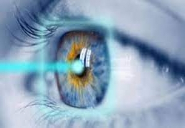
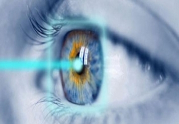
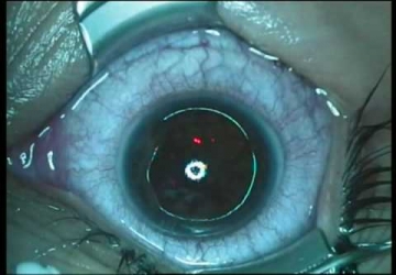
عملية "كيرافليكس" هي عملية لتسطيح القرنية (تقليل تحدب القرنية). تعيد "كيرافليكس" تشكيل القرنية حرارياً من دون إزالة أي نوع من الأنسجة لذلك توفر هذه العملية قدرة فريدة على تصحيح الانكسار من دون إضعاف سلامة النشاط الحيوي للقرنية، كما يحدث مع الليزك وغيرها من عمليات تصحيح الانكسار .
وتقوم عملية "كيرافليكس" بتغير تحدب القرنية المخروطية عن طريق تغيير الضغط في ألياف الكولاجين حرارياً، وتحقق قرنية اقل تحدبا، ويمنح ذلك تصحيح الانكسار وتجانس القرنية غير المنتظمة بالنسبة لحالة القرنية المخروطية، وتالياً تحسين حدة البصر.
فالاستفادة من تشابك عملية "كيرافليكس" هي عملية لتسطيح القرنية (تقليل تحدب القرنية). تعيد "كيرافليكس" تشكيل القرنية حرارياً من دون إزالة أي نوع من الأنسجة لذلك توفر هذه العملية قدرة فريدة على تصحيح الانكسار من دون إضعاف سلامة النشاط الحيوي للقرنية، كما يحدث مع الليزك وغيرها من عمليات تصحيح الانكسار .كولاجين القرنية يحسن استقرار القرنية ويعمل أيضاً على وقف تطور مرض القرنية المخروطية وهذا هو الجديد على صعيد علاج القرنية المخروطية والذي سوف يحدث ثورة في عالم طب العيون لعلاج مرض القرنية المخروطية.
Read More..
محاذير قبل اجراء تصحيح النظر
قبل إجراء عملية تصحيح النظر بالليزر، يجب أخذ مجموعة من الإحتياطات الهامة لتفادي أي آثار جانبية يمكن أن تصيب العين، إليك التفاصيل
Read More..

الزيغ البصري
يشير الانحراف في البصريات إلى وجود عيب في العدسة بحيث لا يركز الضوء على نقطة ما ، ولكنه ينتشر في بعض مناطق الفضاء ، وبالتالي فإن الصورة التي تكونت بواسطة عدسة ذات انحراف غير واضحة أو مشوهة ، مع طبيعة تشويه اعتمادا على نوع من الانحراف.
Read More..
تصليب القرنية
هو علاج ضوئي كيميائي، يهدف إلى تماسك القرنية وزيادة صلابتها، وهو العلاج الأحدث والأكثر أماناً لعلاج القرنية المخروطية المبكرة.
Read More..
Vision correction operations
عمليات تصحيح الإبصار
عملية تصحيح النظر بالليزر لإزالة النظارات اكثر سهولة الآن من أي وقت مضى. ومع ذلك، فان هناك طرقا مختلفة لتنفيذ جراحة ازالة النظارات بالليزر. ليست كل طريقة مناسبة للجميع، لذلك يجب اختيار النوع الاكثر ملاءمة لكل حالة
وبسؤال الدكتور محمد حنتيره _استاذ مساعد شرفى _قسم العيون _جامعة أم القرى _السعودية
وضح لنا بعض عمليات تصحيح الإبصار
عملية تصحيح النظر بطريقة ASA
وفق طريقة ASA (عملية إزالة الخلايا المتقدمة من السطح ) يتم العلاج في الجزء الخارجي من القرنية دون أي جروح أو أي إجراء جراحي اخر. تتميز هذه الطريقة بفترة الشفاء التي تستغرق عدة أيام والشعور بعدم الراحة خلال هذه الأيام. ومع ذلك، فان هذه الطريقة تعتبر امنة وبسيطة
عملية تصحيح النظر بتكنولوجيا الـ Z-LASIK
إزالة النظارات بهذه التكنولوجيا تقلل إلى الحد الأدنى من المخاطر وتؤدي إلى الشفاء السريع
في طريقة Z-LASIK، تشكل ومضات شديدة الدقة من أشعة الليزر سطحا من الفقاعات الصغيرة التي تفصل الطبقة العليا من القرنية وتسمح بإنشاء نافذة علاجية. طريقة الـ- Z-LASIK هي في الواقع تطوير لعملية اللاسيك العادية التي لا تزال تستخدم ، والتي تستخدم فيها سكينا، لذلك فهنالك مخاطر وعدم دقة، بالمقارنة مع طريقة الـ- Z-LASIK الأكثر نجاحا، حيث يتمكن المريض من الرؤية بشكل جيد على الفور بعد العلاج من دون ألم وبقليل من الاثار الجانبية
• مزايا عملية تصحيح النظر أو إزالة النظارات بالليزر بواسطة Z-LASIK:
تأثير الـ Z-LASIK على العين صغير جدا. هذه العملية تستغرق دقائق قليلة لكل عين. الوقت الذي يلمس فيه شعاع الليزر العين قليل إلى أدنى حد ممكن
طريقة Z-LASIK يسمح بإجراء جراحة إزالة النظارات أيضا للأشخاص الذين تبين إنهم غير ملائمين لعملية لاسيك في الماضي، بمن في ذلك الأشخاص الذين يعانون من قصر النظر الشديد أو ذوو القرنية الرقيقة من المتوسط
هي طريقة مريحة وغير مؤلمة: لا يتم تثبيت رأس المريض خلال فترة العلاج
فترة الانتعاش قصيرة بشكل كبير، بالمقارنة مع الطرق الأخرى. عادة، يمكن للمريض أن يرى جيدا في غضون بضع ساعات بعد الجراحة
اثار جانبية اقل: طريقة Z-LASIK تسمح للجراح بتوسيع مساحة العلاج. ولذلك يقل خطر حدوث الاثار الجانبية غير المرغوب فيها، مثل جفاف العينين واضطرابات الرؤية الليلية
ومع ذلك، فان طريقة الـ Z-LASIK ليست مناسبة لكل شخص، تكلفتها أكبر من تكلفة عملية الـ ASA ، كما يمكن أن يشعر الشخص المعالج بالجفاف لفترة قصيرة بعد العلاج
عملية تصحيح النظر بواسطة زرع حلقات القرنية بطريقة FERRARA
هذا هو الحل الذي يوصى به للأشخاص ذوي القرنيات غير الطبيعية، ولأولئك الذين يعانون من تمخرط القرنية أو اللابؤرية الشديدة
يتم إدخال حلقات رقيقة غير مرئية إلى داخل القرنية، حيث تتغير درجة تقعرها وتمنحها المرونة التي تحسن الرؤية فورا. وثمة ميزة أخرى لهذه الطريقة هي أنها طريقة قابلة للعكس، عن طريق إزالة الحلقات أو استبدالها.
عملية تصحيح النظر بواسطة زرع عدسة داخل العين
في الحالات التي يكون فيها قصر النظر شديدا (أكثر من 10، عادة)، أو بعد النظر شديدا (أكثر من 5 +)، أو قرنيات رقيقة جدا، يمكن زرع عدسة تحت القرنية في الجزء الأمامي من العين (الغرفة الأمامية) أو في الجزء الخلفي من العين (الغرفة الخلفية). هذا العلاج يكون مناسبا للأشخاص الذين يستخدمون نظارات ذات درجات مرتفعة، لأنه يسمح بالحفاظ على سلامة القرنية وقوتها، كما أنه قابل للعكس، ويمكن إزالة العدسة أو استبدالها إذا لزم الأمر
فحص ملاءمة عملية تصحيح النظر
في كل الأحوال، ينبغي قبل الخضوع لعملية جراحية لإزالة النظارات بالليزر، وبأي طريقة كانت، إجراء فحص الملاءمة. يتم إجراء فحص الملاءمة الكاملة من قبل فريق من الخبراء يتألف من طبيب العيون وطبيب اختصاصي. ينبغي أن تعطى أفضلية للحالة التي يكون فيها الجراح هو نفسه الذي يجري فحص الملاءمة
Laser vision correction for eyeglass removal is now easier than ever. However, there are various ways to perform laser eyeglass removal surgery. Not every method is suitable for everyone, so the one that is best suited for each situation must be chosen
Read More..
Imaging for visual coherence
Coherence imaging is an imaging examination of the inner tissues of the eye - the network and the optic nerve.
Read More..
Imaging for visual coherence
Imaging for visual coherence
Coherence imaging is an imaging examination of the inner tissues of the eye - the network and the optic nerve.
With the question of Dr. Muhammad Hantira - Honorary Assistant Professor - Ophthalmology Department - Umm Al-Qura University - Saudi Arabia
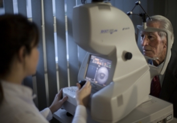
Explain that drops are used to dilate the pupils, and then the person sits opposite a device that sends a beam of light into the eye, and the return waves are measured from the fabricated tissues (similarly to ultrasound).
Depending on the time it takes for these waves to return, an image of the inner components of the eyeball is created and color images of different tissues are created.
The thickness of different tissues - such as nerve tissue - can also be measured in order to track changes stemming from different diseases.
Warnings
• Public
No, except for the side effects associated with dilated pupils (blinding, blurred vision lasts up to 4 hours).
• Driving:
It is not possible to drive up to 4 hours of the examination due to dilated pupils.
Examination results
• In men
Sound results:
In a healthy eye, a healthy retinal bottle and optic nerve template are obtained.
• In women
Sound results:
In a healthy eye, a healthy retinal bottle and optic nerve template are obtained.
• In children
Sound results:
In a healthy eye, a healthy retinal bottle and optic nerve template are obtained.
Read More..
Laser vision correction operations
Laser vision correction operations
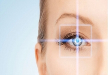
They are operations in which the curvature of the cornea is controlled by a laser in a way that treats myopia, farsightedness and astigmatism, which ultimately leads to the formation of a clear image on the retina, and the complete dispensing of medical glasses and contact lenses.
What are the defects of vision?
• Myopia is the inability of the eye to see distant objects clearly.
• Farsightedness: It is the inability of the eye to see near and far objects without eye fatigue.
• Astigmatism: Astigmatism occurs as a result of irregularities in the surface of the cornea of the eye, which leads to headache and blurred vision.
How can vision defects be corrected?
• Lasers: all kinds (regular, femtolysk, castm lasik, surface laser, femtosil).
In all of them, the curvature of the cornea is controlled in a way that addresses myopia, farsightedness and astigmatism with great effectiveness. LASIK is not a surgical procedure. Rather, it is a laser correction of corneal curvature in a few seconds, and its effect lasts for life in most cases.
• Lens implantation (ICL):
It has witnessed a great development in recent years, especially after the emergence of the last generation of lenses implanted in the posterior chamber of the eye, which increased the degree of safety and effectiveness of the operation by nearly 100% and made the operation the ideal solution for patients who suffer from weak cornea or severe nearsightedness.
• The latest laser vision correction procedures
1- Regular LASIK.
2- Detailed LASIK.
3- Femto-Lysic.
4- Femto-custom lasik.
5- Femto Smile.
How is the patient prepared before LASIK?
• Before LASIK, the doctor examines the eye in detail to determine the following with the utmost accuracy:
• Accurately know the degree of nearsightedness or farsightedness, and accurately determine the degree and axis of associated astigmatism.
• Ensure that the eye is free from diseases such as high eye pressure, cataracts, retinal diseases, and corneal clouds.
• Making a topographic drawing of the cornea with the pentagonal camera, which imaging the topography of both the front and back surfaces of the cornea and determining the thickness of the cornea with the utmost accuracy, then after that the image is analyzed by computer to find out the regularity of the corneal surfaces and make sure that they are free of keratoconus.
• Creating a map of the visual distortions of the eye and taking an eye print using a technology that identifies the problems that the optical system of the eye suffers from, which leads to a lack of efficiency of night vision even using glasses, then the computer attached to the LASIK device is fed with the necessary data in order to detail LASIK taking into account the treatment of these problems, This is called detailed LASIK or Alcastum LASIK.
What steps take place inside the operating room and the time taken?
• The operation does not take more than 5 minutes, the operation is performed under the influence of local anesthesia, and the patient does not feel any pain during the operation, and then feels a slight burning sensation for a period of 2 - 6 hours.
• All that is required of the patient is not to move and respond to some of the instructions that the doctor gives him during the operation, then the patient moves to the waiting room until the doctor examines him again and the patient returns home.
Read More..
Keratoplasty
Keratoplasty
Keratoplasty procedure is used in the event that there is an abnormality in the cornea of the eye, what does corneal fixation mean and how is this process performed, here is this article for the details of the corneal fixation process.
Keratoplasty is an outpatient procedure used to treat keratoconus and some other conditions that may cause a weakening of the cornea.
And with the question of Dr. Muhammad Hantira - Honorary Assistant Professor - Department of Ophthalmology - Umm Al-Qura University - Saudi Arabia
Explain that the cornea is stabilized by creating new collagen fibers within the cornea, which leads to a better stabilization of the cornea.
This procedure is done with riboflavin, known as vitamin B, followed by ultraviolet light.
Types of keratoplasty
There are two basic types of corneal fixation, they are:
• Fixation of the cornea by removing the epithelium
It is done by removing the thin outer layer of the cornea, known as the epithelium, which facilitates the passage of riboflavin through the corneal tissue.
• Fixation of the cornea without removing the epithelium
This type is called corneal epithelial fixation.
How to pre-evaluate the corneal fixation?
Dr. Hantereh evaluates the patient before performing the corneal fixation procedure, to ensure the patient's readiness for the operation.
The doctor measures corneal thickness, visual acuity, and eye health in general, and in addition, the doctor determines the condition of the eye to ensure the patient is ready for this procedure.
What are the procedures for corneal fixation?
The corneal fixation process takes place in Dr. Hantereh's clinic, as he begins by applying numbing drops to the eye and then applying drops of riboflavin, in order to facilitate better absorption of light from the cornea, as the absorption process takes approximately half an hour.
After that, light is shed on the eye, and it is worth noting that the procedure is painless, especially with the presence of anesthesia, and it should be noted that the operation takes from an hour to an hour and a half only.
What are the instructions that must be followed after the corneal fixation?
There are some procedures that the patient must do after the corneal fixation procedure, as these procedures include the following:
• Using antibiotic drops and steroid drops, as they remain for about a week after the operation.
• Using artificial tear drops for relatively long periods of time after the procedure.
• Putting contact lens dressings in the eye, in order to ensure the comfort of the eye, and it should be noted that these dressings should be replaced if they fall out.
• To see the doctor continuously in the first period after the operation.
• Be careful not to rub the eyes for five days after the operation.
• Wearing sunglasses, especially when you have photosensitivity.
• See a doctor immediately if there is severe pain or some unexplained symptoms.
What are the complications that may result from the corneal fixation?
Some complications may occur as a result of the corneal fixation process, as these complications include the following:
• Destruction of the cornea or external epithelium.
• Eye swelling or pain.
• Infection in the eye.
• Vision problems, such as blurry vision.
Read More..
Photochemotherapy
Photochemotherapy is considered one of the most recent treatment methods that increase the mechanical and biochemical tolerance of the collagen that contains the corneal fibers.
Read More..
fluorescein
Fundus imaging after fluorescein injection
This is an examination in which the fundus is visualized after an intravenous fluorescein (a contrast agent) is injected.
And with the question of Dr. Muhammad Hantira, Honorary Assistant, Ophthalmology Department, Umm Al-Qura University, Saudi Arabia
Explanation of this examination gives information about physical or functional changes in the retina, choroid and optic nerve.
How is filming done?
Dr. Hantira explained:
After the pupil is dilated with drops, a fluorescein-type contrast agent is injected into the vein. The contrast material is concentrated in the blood vessels in the retina and in the choroid. The person to be examined sits in front of a special camera to photograph the fundus (the back part).
Imaging is done several times at specific intervals before and after the fluorescein is injected.
Warnings
• Public
The risk is very small. Side effects may occur: Due to dilatation of the pupils of the eyes for the purpose of the examination: Blurred vision may occur for a period of up to 4 hours after the examination. (Difficulty driving, difficulty reading). And because of fluorescein: the skin and conjunctiva may become brownish-yellow, which will disappear after 6-12 hours. The urine may be brownish-yellow and disappear after 24 hours. The listed side effects are harmless and do not involve any risks. Nausea, vomiting, emaciation and feeling dizzy - these effects may occur shortly after the examination, and then disappear without causing any harm. Redness or soreness may occur at the injection site. Rash and breathing difficulties rarely appear, until an allergic (allergic) reaction reaches anaphylaxis. In very rare cases (one in 250,000 people who undergo this test) a heart attack may occur. However, this examination is generally safe and does not pose any risks.
• During pregnancy:
There are no warnings.
• Breast feeding:
There are no warnings.
• Babies and children
There are no warnings.
• Elderly:
There are no warnings.
• Driving:
Do not drive for 4 hours after the test, because the pupils have dilated.
Examination results
• In men
The right results:
There is a specific structure to the image. When an abnormal result appears, the doctor will analyze it according to imaging results, eye examination results, medical history, etc.
• In women
The right results:
There is a specific structure to the image. When an abnormal result appears, the doctor will analyze it according to imaging results, eye examination results, medical history, etc.
• In children
The right results:
There is a specific structure to the image. When an abnormal result appears, the doctor will analyze it according to the imaging results, the results of the eye examination, the medical history, etc. (It is rarely necessary to perform this examination in children
Analysis of the results
Disorders of the blood vessels of the retina
It may result from one or more of the following disorders:
• Retinopathy due to diabetes
• Retinopathy due to hypertension
• Macular degeneration
• Optical papilledema
• Retinal tear
• Retinitis pigmentosa
Read More..
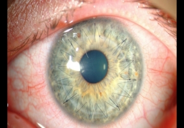
Corneal transplant
Corneal transplant
Operation goal:
The purpose of performing a corneal transplant is to replace the affected cornea of the eye, due to its exposure to many factors, which can lead to permanent and irreparable diseases or damage. The cornea for transplantation is taken from a dead donor, while this is the most common procedure among various organ transplants.
With the question of Dr. Muhammad Hantira, an honorary assistant professor, ophthalmology department, Umm Al-Qura University, Saudi Arabia
About the methods used to perform corneal transplantation stated:
There are many methods used for corneal transplant surgery. The old method involves completely removing the affected cornea and stabilizing the healthy cornea with sutures (sutures). But when using modern methods that use precise devices such as lasers, only part of the cornea (DSEK) is removed. Also, there are other types of corneal transplant procedures.
Prepare for the operation:
There is no need to match the tissue and blood type when performing corneal transplantation surgery, because there are good treatments that are given to the patient after transplantation, which contribute to the acceptance of the transplanted organ with a high success rate of about 90%. Another reason why such a fit is not needed is that the cornea is a tissue that is not attached to a blood vessel.
In most cases, there is no need for a general examination before surgery. The ophthalmologist performs a comprehensive examination of the eyes, which includes checking the extent of vision, pressure inside the eyes, visual acuity and examining the fundus, in order to ensure the integrity of the retina.
The cornea is transplanted under local or general anesthesia. The doctor should also be consulted with regard to all medications that the patient should stop taking before the operation date. Also, there is a need to fast for 8 hours before the operation.
process:
After instillation of the dilating material into the pupil, local anesthesia is administered to the area or general anesthesia if necessary. According to the traditional method, the surgeon makes a small, thin incision in the cornea, and then removes all corneal tissue. After that, the surgeon sutures the transplanted cornea in its place.
In modern methods of corneal transplantation surgery, there is not always a need to transplant the entire cornea, and only the lining of the cornea can be transplanted (if it is injured) or another part of the cornea is implanted using accurate and modern devices. In laser-based surgery, the cornea that will be transplanted is cut according to the required size and the extent of damage to the original cornea. Then they are fixed - sometimes without the need for stitches -. At the end of the operation, a bandage is placed over the eye.
The surgery takes one to two hours (depending on the type of surgery).
Post-operative treatment:
After corneal transplant surgery:
The patient stays in the hospital under observation for one or a few nights. When modern surgical methods are used, patients are sometimes able to leave the hospital earlier. The patient is instructed on how to use eye drops that contain antibiotics to prevent infection, which he should use for 3 weeks after the operation.
The patient is provided with eye drops to prevent eye infection and to suppress the immune system.
The patient can take pain relievers as needed. While the recovery period is related to the type of corneal transplantation that was followed, as this period is short when using modern methods.
The sutures (sutures) are removed if they are used in surgery, after a few months have passed (in traditional corneal transplant surgery).
In the following cases, the patient should refer directly to the doctor: persistent pain despite the use of analgesics, secretions from the eye, bleeding from the eye, or a high temperature.
Read More..
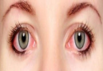
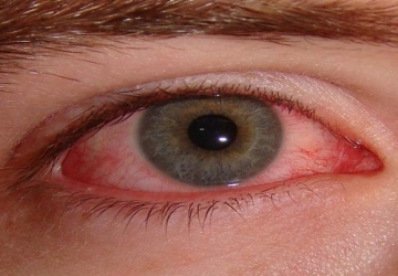
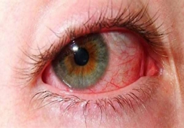
Redness of the eye
Six surprising causes of red eyes, most notably stress and smoking
Dr. Mohamed Hantira _ Honorary Assistant Professor mentioned
Ophthalmology Department - Umm Al-Qura University _ Saudi Arabia 6 surprising causes of redness and blood congestion in the eye, and these reasons include the following:
Focusing a lot while working and looking for long periods at the computer screen or when using a mobile phone, this habit can cause dryness and eye irritation, which results in blood congestion in the eyes.
Seasonal allergies can cause red eyes, because there are some allergens present in the environment such as pollen that can lead to inflammation of blood vessels in the eyes.
Some medicines If you are taking some strong medicines such as anti-anxiety drugs and antibiotics, they can cause dry eyes, making them appear bloodshot.
Smoking can cause dry eyes, and lead to inflammation of the blood vessels in the eye, which causes the eyes to become infected with blood.
Swimming Some people may experience eye congestion with blood, especially after swimming, whether in swimming pools or the sea, as a result of exposure to chlorine and salts that result in irritation and congestion in the eyes.
Stress and tension can cause eye congestion, because stress increases blood flow to the brain and optic nerves.
Read More..
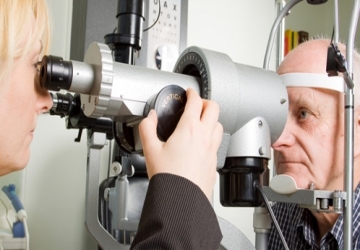
Fundoscopy
Fundoscopy
In the fundoscopy test, the fundus is examined. With a special device (called a fundus endoscope), the doctor looks through the pupil of the eye at the retina, the optic nerve, the retinal blood vessels and the inner cavity of the eye. The retina forms the back of the eyeball. In fact, this is the only place in the body where blood vessels can be seen as they are.
How to take the test
With the question of Dr. Muhammad Hantira, an honorary assistant professor, the Eye Department, Umm Al-Qura University, Saudi Arabia
Explain that the eyeball can be seen directly or indirectly.
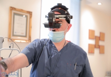
In the first method, the doctor looks with an ophthalmoscope that lights the retina directly or through a crack lamp. In the first method, the doctor places a mirror on his head that reflects light inside the retina and looks at the inside of the eyeball, using a magnifying lens.
With the help of this examination, which lasts about 5-10 minutes, it is possible to see damage to the eye itself, or to notice systemic diseases that also appear in the retina.
Warnings
• Public
There are no risks.
• During pregnancy:
There are no special effects or problems.
• Breast feeding:
There are no special effects or problems.
• Babies and children
There are no problems or special effects. The patient's cooperation is needed, and so the examination may be difficult. When examining young children and infants to assess the need for glasses or to diagnose strabismus, the examination should be performed when the pupils are dilated. Sometimes, a stronger distillation of a substance (such as cyclopentolate or atropine) is needed in young children. Some of the children have red cheeks, fever, rash, or a rapid pulse.
In rare cases - drowsiness or hyperactivity, sometimes even convulsions.
• Elderly:
There are no special effects or problems.
• Driving:
The subject may suffer from blurred vision and dazzling that lasts for several hours after the examination, as a result of the dilated pupil.
Medicines that affect the test result
Medicines that affect the autonomic nervous system, such as adrenaline, atropine, beta-blockers and others, may cause a permanent change in the size of the pupil, but they do not affect the retina. Therefore, some of these drugs may also make it difficult to perform the test, but they do not affect the results.
Analysis of the results
• High intracranial pressure
• Optic neuritis
• White reticulum similar to milk:
• Severe retinal artery blockage
• Narrowing of blood vessels
• high blood pressure
• Extensive retinal hemorrhage:
• Severe retinal vein blockage
• Retinal detachment
• A tumor in the retina
• White, cotton-like patches
In the fundoscopy test, the fundus is examined. With a special device (called a fundus endoscope), the doctor looks through the pupil of the eye at the retina, the optic nerve, the retinal blood vessels and the inner cavity of the eye. The retina forms the back of the eyeball. In fact, this is the only place in the body where blood vessels can be seen as they are.
Read More..
Smoking and eye health
Smoking and eye health
Dr. Muhammad Hantira, an honorary assistant professor, the Ophthalmology Department, Umm Al-Qura University, Saudi Arabia, said that smoking is one of the most harmful habits for the eyes, as smokers are considered four times more likely to be blind with age, according to a study of the British Medical Journal.
Dr. Hanatera explained that smoking leads to serious eye problems, including.
Dry eye syndrome
Dry eye syndrome is a common condition that occurs when the eye does not lack enough or the right kind of tears. Cigarette smoke is known to contain more than 7,000 chemicals, and they can cause irritation and damage during dry eye syndrome.
Symptoms of dry eye syndrome include a burning sensation, itchy eyes, redness, a sandy sensation, and severe irritation.
Experts prone to dry eyes recommend avoiding smoking and contact with smoke. Because smokers are twice as likely to develop Dry Eye Syndrome.
Cataract
Cataract is a cataract that affects the eye's natural lens. Cataract causes blurry vision and makes colors appear faded, faded, or yellowish. Cataract is the leading cause of blindness and moderate visual impairment worldwide.
If you smoke, you are more likely to develop a cataract, as smoking changes the cells of the lens of the eye through oxidation, which helps the accumulation of heavy metals in the lens, which in turn leads to the formation of cataracts in the eye and its cataract.
Macular degeneration due to age
Age-related macular degeneration occurs when a portion of the retina, called the macula, is damaged. The patient loses central vision and cannot see fine details, although peripheral or side vision is healthy.
In some cases, medical treatments can help reduce the complications of age-related macular degeneration, but there is still no cure.
Although age is the first risk factor in disease, smoking is second. Smokers are more than four times more likely to have this condition compared to non-smokers. Those who live with smokers are twice as likely to develop this condition as well.
Dr. Huntereh noted that recent medical advances have introduced treatment for advanced cases of macular degeneration in the form of an injection into the eye to restore vision. Research has shown that smokers are less likely to respond to these injections compared to non-smokers.
Diabetic Retinopathy
Smokers with diabetes are also at risk of developing diabetic retinopathy, which is the damage to the blood vessels in the eyes.
Smoking causes blood vessels to narrow, which reduces blood flow to the eyes.
This can lead to diabetic retinopathy, which is characterized by symptoms such as blurred or distorted vision, leading to blindness.
Additionally, smoking raises blood sugar and can make the body more insulin-resistant, which may lead to high blood sugar levels.
Uncontrolled blood sugar can lead to serious complications, including diabetic retinopathy.
Smoking is the only controllable risk factor that contributes to the development of many eye diseases.
Dr. Mohamed Hantira advises patients to participate in programs to support them on their journey to kick this harmful hbit.
Not only does quitting smoking reduce the chance of developing eye problems, but it also helps improve their overall quality of life.
Read More..
Diagnosis and treatment of eye allergies
Diagnosis and treatment of eye allergies
With the question of Dr. Muhammad Hantira, an honorary assistant professor, ophthalmology department, Umm Al-Qura University, Saudi Arabia
Explain that: -
You can get the appropriate diagnosis by referring to an ophthalmologist or an eye allergy specialist, who often comes out with the diagnosis after inquiring about the symptoms and taking a tissue sample in an examination called (the skin prick test).
As for treating sensitive eyes, there are many options and methods, and these are the most important of them:
1- Eye drops
There are over-the-counter types of eye drops available in pharmacies to suit different eye allergies.
It is possible to resort to eye drops that contain active ingredients to combat allergies, or only to use eye drops that work to moisturize the eyes (or the so-called artificial tears).
And since some types of eye drops are not suitable for everyone and may even cause allergic reactions, you must always consult your doctor first about the types most appropriate to the patient's condition.
2- Medicines
There are many medications available that can significantly reduce the symptoms of eye allergies, such as: antihistamines, decongestants, and steroids.
3- Allergy injection
In very severe cases of eye allergy, which other treatments may not work with, it is possible to resort to allergy shots, through which the body is injected with allergens with a gradual increase in the dose each time to improve the body's reaction to these causes.
Cases similar to eye sensitivity
Dr. Hantereh indicated: -
There are cases that may appear at first glance very similar to eye allergy, but they are in fact completely different cases, and they must be dealt with in a different way, and these are some of them:
• Dry eyes.
• Conjunctivitis caused by a virus or bacteria.
• Blockage of the tear ducts.
Read More..
he difference between dry eyes and allergic eyes
he difference between dry eyes and allergic eyes
With the question of Dr. Muhammad Hantira, an honorary assistant professor, ophthalmology department, Umm Al-Qura University, Saudi Arabia
We explained the difference between dry eyes and eye sensitivity. Eye sensitivity, also known as allergic conjunctivitis, is a very common condition, and it occurs when the eyes are affected by something irritating, as the eye secretes a substance called histamine to fight allergens, and as a result the eyelids and conjunctiva become red and swollen.
Tears may flow from the eyes with a burning feeling, and unlike other types of conjunctivitis, eye allergies do not spread from one person to another, and it may be a temporary condition associated with seasonal allergies.
As for dry eyes, it occurs when the eyes are not able to provide adequate moisture to the eyes, and dry eyes can occur if the eye does not produce enough tears, or if it produces poor quality tears, and the person may feel a stinging or burning sensation in the eyes.
Dry eyes may occur in certain cases, such as: riding an airplane, being in an air-conditioned room or when riding a bike, or after looking at a computer screen for a few hours, and eye drops help relieve pain.
Dr. Hantira also indicated that the symptoms differed between them.
Eye allergy symptoms You may suffer from allergic reactions in one eye or both eyes. Symptoms of eye allergy include the following:
• Watery eyes.
• Itching.
• Sensitivity to light.
• Redness.
• The feeling of having sand in the eye.
• Eyelid swelling.
Dry eye symptoms Common dry eye symptoms include:
• Burning and itching.
• Pain or redness.
• Mucus in or around the eyes.
• Periods of watery eyes followed by dry eyes.
• Inability to cry.
• Blurred vision, or eye fatigue.
• Sensitivity to light.
• Heavy eyelids.
• Difficulty wearing contact lenses.
• Eye strain from reading or using the computer.
Read More..
التهاب القزحية
veitis is a type of inflammation that affects the eye, as the iris is known as one of the parts of the middle layer of the eye known as the uvea.
Symptoms and signs of uveitis
With the question of Dr. Muhammad Hantira, an honorary assistant professor, ophthalmology department, Umm Al-Qura University, Saudi Arabia
Explain that there are many symptoms associated with uveitis, including:
•Severe eye pain when exposed to bright light.
• Low vision or blurred vision.
•Pain in the eyes and eyebrow area.
•Redness, especially around the iris.
•Change in the shape or size of the pupil.
•a headache.
Causes of uveitis
Dr. Hantereh explained
Uveitis occurs for unknown reasons, and in these cases the inflammation appears only once.
But many causes may contribute to uveitis, and most of the time the inflammation occurs repeatedly, as the causes of uveitis include the following:
•Eye injury.
•Inflammation of eye surgery.
•Infections such as tuberculosis virus, herpes virus, and others.
•Sarcoidosis.
•Behçet's disease (Adamantiades-Behçet's disease).
• The use of some medications, such as those used to treat glaucoma.
•Autoimmune diseases, such as seborrheic fever and rheumatoid arthritis.
Risk factors for uveitis
Dr. Hantereh indicated:
There are many factors that increase the risk of developing uveitis, as these factors include the following:
•Smoking. Smoking increases the risk of developing iritis.
• Having a sexually transmitted infection, such as AIDS.
•Immune disorders or a weakened immune system.
•Genetic changes.
Diagnosing uveitis
Dr. Hantira explained:
An ophthalmologist diagnoses uveitis, who performs a complete eye examination, and this examination includes the following:
•External examination of the eye.
•Slit lamp examination.
•Visual acuity test.
Treating uveitis
Dr. Hantereh noted:
It is imperative to begin treatment for uveitis immediately upon confirmation of the diagnosis, and it may require more than one follow-up visit to the doctor to finish the treatment.
It is worth noting that there are some medical treatments used for uveitis, which include the following:
•Steroid: Steroid is used to reduce inflammation, as the doctor begins by prescribing steroid eye drops, and if the patient does not respond within a week of treatment with them, he can prescribe pills or needles.
•Eye drops: Eye drops are used to treat uveitis, to help dilate the pupil and prevent contractions.
•Antibiotic or viral: In cases of bacterial infection, antibiotics and antivirals are used in the event of viral infections in the iris.
• Anticholinergic drugs: Anticholinergics reduce pain and light sensitivity by blocking the nerve signals for each.
Prevention methods for uveitis
Dr. Hantereh indicated:
Noting that there are many methods used to prevent uveitis, as these methods include the following:
• Avoid practices that increase the risk of developing iritis, such as smoking.
•Protect the eye from injuries.
•Reducing the eye's exposure to infection.
•Eat healthy diets rich in vitamins, such as vitamin C.
•Do a stress-free exercise, such as yoga.
Read More..
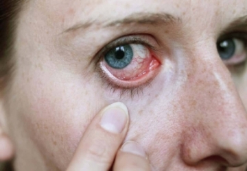
Eye redness
Eye redness The redness of the eye means the appearance of the white of the eye (sclera) in a red color, and is most often caused by inflammation ofthe conjunctiva, expansion of blood vessels in it, or as a result of dry eyes, or exposure to infection, and redness of the eye may be a sign of a medical emergency in some Cases, so it is advised not to neglect the redness of the eye, and to see a doctor to diagnose the condition, especially if the redness of the eye persists for a long time.
Causes of redness of the eye Causes of redness of the eye include:
• conjunctivitis
• Computer vision syndrome
• Eye injuries
• Corneal ulcers
• Eye herpes
• Dry Eye
• Sensitivity
• contact lenses
• Iritis
• Glaucoma
• Excessive use of eye drops to bleach
• Cold and flu
• Hormonal changes during pregnancy
• Smoking and alcohol use
• Exposure to environmental factors such as dust and smoke
• swimming
Symptoms that necessitate seeing a doctor You should see a doctor if eye redness is accompanied by the following symptoms:
• Inability to keep an eye open.
• Eye pain and fever.
• Nausea or vomiting.
• Sudden blurred vision.
• Eye swelling.
• Sensitivity to light.
• Seeing halos around lights.
• Severe headache.
• Noticing a foreign object in the eye.
Treatment of redness of the eye To get rid of red eyes, you can follow the following tips:
• Put warm compresses on the eye for ten minutes to increase blood flow to the eye, and increase the secretion of oils to help soften the eye.
• Applying cold compresses: If the eye does not respond to hot compresses, cold compresses can be used to relieve eye irritation and swelling.
• Artificial tears: They are used to treat redness resulting from dry eyes, in addition to their role in cleaning the eys.
• If the redness is caused by the use of contact lenses, you can consult a doctor and replace the contact lenses with suitable ones, and choose good quality lens preservation solutions.
Drink enough water, as a person needs to drink (8) cups of water; To maintain the fluid balance in the body.
• Limiting the intake of foods that cause inflammation and that may increase the redness of the eye, such as: fast food, dairy products, and processed foods.
• If the eye condition does not improve, a doctor should be consulted, who may prescribe anti-allergies and antibiotics.
Read More..
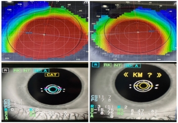
Corneal imaging
Corneal imaging
With the question of Dr. Muhammad Hantira - Honorary Assistant Professor, Department of Ophthalmology - Umm Al-Qura University - Saudi Arabia
About the latest devices used in corneal imaging, the following was mentioned:
Pentacam:
It is one of the most important devices that are used in the most important examinations that must be performed before vision correction (LASIK) operations at Middle East Eye Hospital.
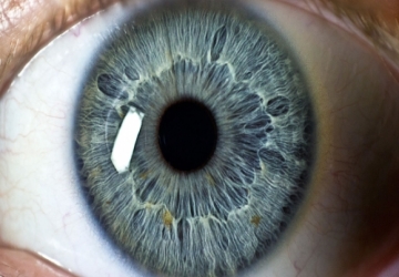
The Pentacam device measures the characteristics of the cornea, as it takes 25 internal and external images of the eye and provides a complete analysis of the cornea and lens of the eye to ensure that the eye health is in the normal range and also determines the most appropriate operation for the patient and helps in early detection of keratoconus for many patients and following its develpment.
Corneal elasticity testing device: Ocular Response Analyzer Hysteresis
This device made a quantum leap in the world of ophthalmology, this device works to determine the flexibility and rigidity of the cornea and thus to know the extent of its acceptance for any kind of vision correction and is considered a complement and support for the diagnostic devices in the Middle East Eye Hospital. In addition, it is one of the most important devices required for patients suffering from high eye pressure, which shows the response of the eye to the drugs used to reduce intraocular pressure and the effectiveness of this treatment on the eye.
Corneal Topography: TopolyzerWavelight T-Cat
Accurate device to diagnose corneal topography using the Placido Disk System, the device returns 22,000 Pex landmarks. To create a topographic map of the eye and then instantly identify visual defects, including deviation, corneal keratosis, astigmatism and tilt.
Even in the most difficult situations. The WaveLightTopolyzer VARIO is the ideal tool for preparing for laser and wave vision correction with precision to the topography of the cornea.
Read More..
Epi Lasik
Epi Lasik surgery is one of the vision correction procedures used in
ophthalmology clinics for the purpose of curving the cornea with the help of laser,
and it is very similar to the regular LASIK procedure except that it includes cutting
a very thin part of the epithelium layer on the surface of the cornea and thus may
be suitable for patients who have a thin cornea.
And with the question of Dr. Muhammad Hantira, an honorary assistant
professor, ophthalmology department, Umm Al-Qura University, Saudi Arabia
Explain to us:
One of the advantages of the LASIK procedure that makes it the most preferred
by many eye surgeons is the lack of the need to use alcohol to facilitate the
separation of the epithelium layer in the cornea, as is the case in LASIK, as many
ophthalmologists consider that the alcohol used has toxic effects on the cells of
the epithelium layer. In the cornea, it may delay the recovery period and return
the patient's vision to a normal level.
The duration of treatment is approximately three days.
What is the reason for resorting to the operation?
Many patients may resort to an Epi-LASIK procedure due to one of the following
problems:
Short sightedness. Farsightedness injury. Farsightedness or astigmatism.
To get rid of prescription glasses or contact lenses.
The necessary checks and procedures before undergoing the operation
Before deciding on the possibility of performing an IBD, a patient must undergo
some tests required by the eye surgeon, including:
A comprehensive eye examination that includes measuring some structures
inside the eye in addition to examining the visual level.
Double endoscopic examination.
Quantitative and qualitative examination of the tear film in the eyes.
Corneal topography; To know the strength and shape of the cornea.
Wave front inspection; To measure high gaze aberration.
Corneal thickness measurement.
Accurate pupil size measurement.
Complications of Epi lasik operation may expose the patient to several risks, and
despite the high success rates of LASIK operations, it may carry some
complications that the patient should be aware of and be aware of, even if they
are rare, such as:
Sensitivity to light.
Dry Eye.
Blurred or cloudy vision.
The need to repeat the operation for not achieving the required level of
vision.
Operation procedure steps
The Epi-Lasik process includes three stages:
Preparing for the operation: The patient is given local anesthesia by using
an anesthetic eye drop, and perhaps giving him a sedative intravenously for
more comfort during the operation.
Before that, the patient is asked not to use contact lenses for at least two
days before the operation.
During the operation: Dr. Hantereh uses a spacer to keep the eyelids wide
apart and the eye open to facilitate work on the cornea, then passes a
special micro-device that contains a sharp blade that cuts a very thin
portion of the corneal surface and separates it from the corneal stroma.
And it allows the passage of intermittent flashes of laser radiation that
reach the cornea and work to reshape its surface in about 20 seconds.
After that, a doctor returns the separated layer of the surface of the
cornea, and he may put a very thin lens on it to help it heal quickly, then
apply a few drops of the antibiotic to avoid infection.
After the operation: the patient can leave the surgeon's clinic immediately
after the operation is over, and it is preferable to accompany one of the
acquaintances or relatives because the patient is not able to drive a car.
The recovery period The success rates of the operation depend mainly on
the patient’s compliance with the eye surgeon's instructions, as being
careful to use eye drops and taking other medicines is the most important
thing that may enhance the chances of recovery and help reach the best
results.
Usually, the patient can notice an improvement in the level of vision after
about a week of the operation and reach the best level of the ability to look
well without glasses after about 3 to 6 months.
Read More..
E.eye
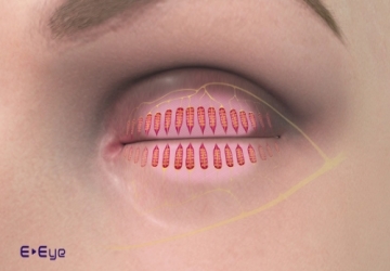
E.eye technique for treating dry eyes
Treatment of dry eyes with modern technology is completely different
from the traditional methods in which dry eyes are treated, in addition
to that it is characterized by the use of a device that takes a sample
from the patient’s tears and examines the proportion of salts in them,
and then an image is measured on the corneal layer to discover the size
of the distribution of the tear in front of the eye.
And with the question of Dr. Muhammad Hantira - Assistant
Professor - Department of Ophthalmology - Umm Al-Qura University -
Saudi Arabia
He mentioned that:
The modern technology for treating dry eyes has proven that among
90% of patients with dry eyes, they do not suffer from problems with
the secretion of tears in the eye, which means that the lacrimal gland is
fully and well functioning, which led to the emergence of this study
recently and the discovery of one of the best modern techniques for
treating dry eyes.
How is dry eyes treated with modern technology?
Dr. Hantira explained:
It is treated in 3 steps, the oil layer is what prevents the tears from
evaporating when exposed to the air, and the movement of the eyelid
is the one that distributes the tears, which prevents the occurrence of
dry eyes and the sebaceous glands that secrete the oily substance into
the tears and are treated and stimulated again to secrete tears to
prevent dry eyes Through the following steps:
1- Stimulating the sebaceous glands:
Treating the glands responsible for secreting tears with modern
technology only takes 3 minutes per eye
The treatment process is done by stimulating these glands and
activating them again to secrete tears and make them appear normal.
2- Use of infrared:
Modern technology for treating dry eyes relies on directing heat to the
glands, making them return to their fluid nature.
3- Nerve stimulation:
The modern technology relies on stimulating the nerves that nourish
the sebaceous glands to modernize, move and activate them in an extra
way and make them perform tasks larger than the normal range until
the dry eye is treated.
Advantages of treating dry eyes with modern technology
• Modern technology works to treat dry eyes and not to compensate
for the lack of sebum secretion
• It does not take long
• After treatment, the patient can return to work normally
• The technology does not have any side effects
• The patient needs only one or two sessions of treatment (severe
cases only need 3 sessions, no more)
• Modern technology treats dry eyes within 5 to 10 minutes (for both
eyes)
Does modern technology treat dry eyes radically?
Treatment with modern technology is radical ... But!
Dr. Hantira explained:
It is necessary to change the lifestyle of everyone who has undergone
treatment with modern technology to treat dry eyes, but what does
this mean!
In a clearer sense, a person who suffers from dry eyes can undergo
treatments, but how is that when he does not drink water in
abundance, which is one of the biggest causes of dry eyes, so at least
every person should drink about two liters of water a day.
It is also not necessary to claim a treatment for dry eyes, despite sitting
in front of the computer for 12 continuous hours
Read More..

Optical coherence tomography (OCT) of the anterior segment
Optical coherence tomography (OCT) of the anterior segment
of the eye.
By asking Dr. Muhammad Hantira, Honorary Assistant Professor,
Ophthalmology Department, Umm Al-Qura University, Saudi Arabia,
about the role of the computerized tomography device in the field of
ophthalmology, he explained to us the following:
Optical coherence tomography (OCT) of the anterior segment of the
eye allows us to obtain high-resolution images from the front of the eye
using a property of light called optical interference. Currently it has
become a very useful tool for studying the eye's microscopic
anatomical structure.
Optical coherence tomography (OCT) of the anterior segment of the
eye uses:
• For follow-up of patients with refractive surgery, intra corneal
parenchyma, corneal transplantation, refractive cataract surgery.
• To follow up on patients who are undergoing surgery for glaucoma
leachate.
• In the field of cataract surgery, optical coherence tomography (OCT)
of the anterior segment of the eye allows accurate analysis of the
structure of the surgical incision, as well as the relationship between
the implanted lens and the posterior capsule.
Optical coherence tomography (OCT) of the anterior segment of the
eye is useful for:
• Analysis and evaluation of tumors and cysts in the front of the eye.
• Conjunctival tumors analysis, and various other diseases such as
corneal dystrophy, degeneration and infections.
• It also allows us to determine the thickness and epithelium of the
cornea.
• Determining the iris-corneal angle, measuring the depth of the
anterior chamber, and assessing the position of the implanted lens
inside the eye.
• In addition to studying the placing of contact lenses on the surface of
the cornea, and studying the condition of the surface of the eye, and it
is also useful for studying dry eyes.
Read More..
Cosmetic damage to the eyes
The wrong use of eye makeup leads to the occurrence of some indispensable symptoms that affect your health and your skin, it leads to eyelashes loss and the occurrence of infections in the eyelids in addition to the appearance of dark circles, so eye makeup should be used in a limited way as possible.
Read More..
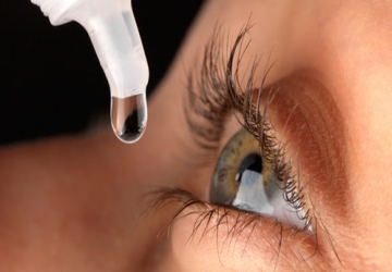
Eye drops
The eye drops are usually the basis of a salt liquid, and the eye drops help to provide the necessary moisture to the eyes or they can be used for purely medicinal and therapeutic purposes.
Read More..
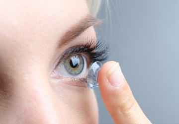
The four best types of contact lenses
The use of contact lenses is no longer a matter for many mothers, as wearing them is not only limited to medical use, but also for cosmetic use, in order to rejuvenate and look their best. These cosmetic lenses are available in green, gray, blue and hazel colors.
Read More..
Epi Lasik
Epi Lasik surgery is one of the vision correction procedures used in ophthalmology clinics for the purpose of curving the cornea with the help of laser, and it is very similar to the regular LASIK procedure except that it includes cutting a very thin part of the epithelium layer on the surface of the cornea and thus may be suitable for patients who have a thin cornea.
Read More..
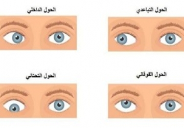
Diagnosis and treatment of strabismus in young and old
Children should be examined by a specialist doctor during childhood stages to detect any possibility of eye diseases. And this matter is important if one of the relatives suffers from the presence of eye squint or laziness.
Read More..
Eye allergy
Eye allergy is a condition that affects the eyes as a reaction, when the eye is exposed to one of the allergens and stimuli, such as dust, pollen or smoke.
Read More..
The operation to enlarge and expand the eyes
That in this article, we will discuss the most important section in eye plastic surgery, which is enlarging the eyes. What is the process, how is it performed, its types, costs, the best places to provide the service, and what you should look for before going through this experience.
Read More..