Fundoscopy
In the fundoscopy test, the fundus is examined. With a special device (called a fundus endoscope), the doctor looks through the pupil of the eye at the retina, the optic nerve, the retinal blood vessels and the inner cavity of the eye. The retina forms the back of the eyeball. In fact, this is the only place in the body where blood vessels can be seen as they are.
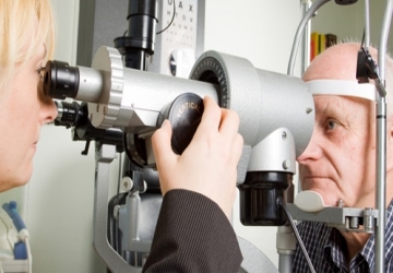
How to take the test
With the question of Dr. Muhammad Hantira, an honorary assistant professor, the Eye Department, Umm Al-Qura University, Saudi Arabia
Explain that the eyeball can be seen directly or indirectly.
In the first method, the doctor looks with an ophthalmoscope that lights the retina directly or through a crack lamp. In the first method, the doctor places a mirror on his head that reflects light inside the retina and looks at the inside of the eyeball, using a magnifying lens.
With the help of this examination, which lasts about 5-10 minutes, it is possible to see damage to the eye itself, or to notice systemic diseases that also appear in the retina.
Warnings
• Public
There are no risks.
• During pregnancy:
There are no special effects or problems.
• Breast feeding:
There are no special effects or problems.
• Babies and children
There are no problems or special effects. The patient's cooperation is needed, and so the examination may be difficult. When examining young children and infants to assess the need for glasses or to diagnose strabismus, the examination should be performed when the pupils are dilated. Sometimes, a stronger distillation of a substance (such as cyclopentolate or atropine) is needed in young children. Some of the children have red cheeks, fever, rash, or a rapid pulse.
In rare cases - drowsiness or hyperactivity, sometimes even convulsions.
• Elderly:
There are no special effects or problems.
• Driving:
The subject may suffer from blurred vision and dazzling that lasts for several hours after the examination, as a result of the dilated pupil.
Medicines that affect the test result
Medicines that affect the autonomic nervous system, such as adrenaline, atropine, beta-blockers and others, may cause a permanent change in the size of the pupil, but they do not affect the retina. Therefore, some of these drugs may also make it difficult to perform the test, but they do not affect the results.
Analysis of the results
• High intracranial pressure
• Optic neuritis
• White reticulum similar to milk:
• Severe retinal artery blockage
• Narrowing of blood vessels
• high blood pressure
• Extensive retinal hemorrhage:
• Severe retinal vein blockage
• Retinal detachment
• A tumor in the retina
• White, cotton-like patches