Imaging for visual coherence
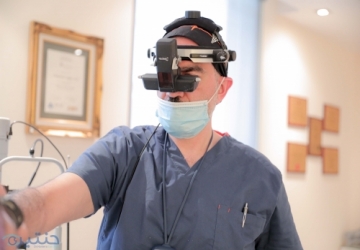
Coherence imaging is an imaging examination of the inner tissues of the eye - the network and the optic nerve.
With the question of Dr. Muhammad Hantira - Honorary Assistant Professor - Ophthalmology Department - Umm Al-Qura University - Saudi Arabia
Explain that drops are used to dilate the pupils, and then the person sits opposite a device that sends a beam of light into the eye, and the return waves are measured from the fabricated tissues (similarly to ultrasound).
Depending on the time it takes for these waves to return, an image of the inner components of the eyeball is created and color images of different tissues are created.
The thickness of different tissues - such as nerve tissue - can also be measured in order to track changes stemming from different diseases.