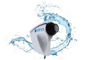
Idra Dry Eye diagnosis
Is the new instrument of individual analysis of tear film that allows to do a quick detailed structural research of the tear composition. Research on all the layers: Lipid, Aqueous, Mucin.
Read More..
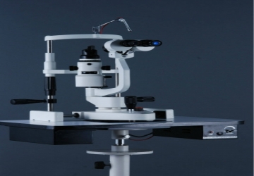
Slit lamp device
Slit lamp device It is a device used to examine the anterior parts of the eye eyelid, conjunctiva, cornea, anterior chamber, lens, iris and anterior portion of the vitreous
Read More..
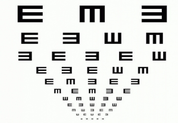
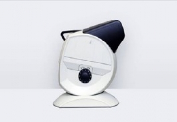
Eye test screen
It is a device used to assess visual acuity by displaying vision test signals on an electronic screen controlled by a remote control.
Read More..
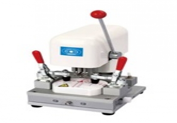
Lens drilling rig
It is a device used to drill a half-frame lens to attach a nylon thread to it. Assist in equipping the half-frame lenses and glasses.
Read More..
Contrast sensitivity check
Contrast sensitivity check
This is a plate used to measure a patient's contrast sensitivity.
the use Measure the patient's contrast sensitivity by noting the last layout that the patient can see during each row of the board.
Read More..
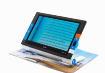
Text magnifier
Text magnifier
It is an electronic device that produces an enlarged image of a text or an object by displaying it on a screen.
Its uses:
• It is used to help the visually impaired to read handwriting in an enlarged way, such as reading newspapers, writing, viewing pictures, and performing close work such as knitting.
Read More..
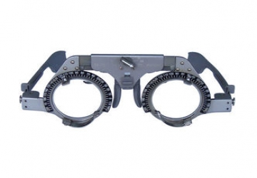
Experimental framework
The experimental frame is a frame used to experiment with lenses, prescriptions and various tests, and its size and dimensions can be changed to suit the patient's face, and the frame contains more than one place for placing the lenses.
Read More..
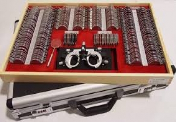
Lens box
It is a set of spherical and cylindrical lenses and prisms placed in a special bag arranged according to the powers and the type of lenses, positive or negative. It also contains a set of accessories.
Read More..
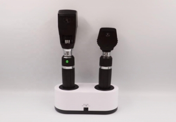
Retinoscope
A portable device used to check the refractive strength of the eye based on observing the movement of the beams bouncing off the retina.
Read More..
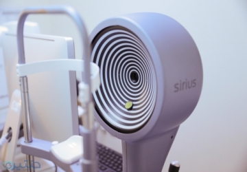
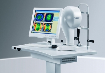
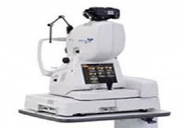
entacam or pentacam examination
It is a very advanced examination performed by a very advanced (CSO Sirius) device, and it is the last science reached in this type of examination as it combines two methods of taking pictures of the cornea, which are the two most accurate and globally widespread methods - the rest of the devices take pictures in one way This greatly enhances the accuracy of the examination.
Read More..

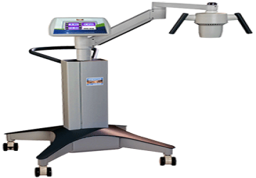
YAG laser to clean the back cover and glaucoma
YAG laser to clean the back cover and glaucoma
Laser is a modern technology that has revolutionized many medical fields, including eye surgery.
Lasers are used in treating cataracts and glaucoma, but the matter differs according to each case, and there are different types of lasers to deal with these problems.
Dr. Muhammad Hantira, an honorary assistant professor at the Ophthalmology Department, Umm Al-Qura University, Saudi Arabia, said
The treatment of cataract and glaucoma on the eye with laser is done in specific cases and varies according to the nature of each case. Cataract uses the femto-cataract technique, which enables us to divide the lens of the eye affected by the cataract with femto-laser.
Hantira added that the use of femto-laser “cataract” in the treatment of cataract achieves good results and facilitates the process, especially in cases of heavy white water, where the laser can get rid of it and treat it completely.
Hantira explained that with regard to glaucoma, we use a type of laser called a “yag laser” through which we can make an opening in the iris of the eye and be able to perform the simple glaucoma operation that affects the patient in its early stages.
Hantira emphasized that severe cases of glaucoma require surgical intervention, and that the “YAG laser” cannot be used to treat them, unlike cataracts, which the “cataract laser” can treat all cases.
A reader asks: My mother suffers from high eye pressure, known as glaucoma, so is the use of lasers useful in treating glaucoma?
Dr. Hantira answers the question, saying:
Glaucoma, known as “glaucoma”, is a disease of high intraocular pressure or pressure forming the eye, and the eye has a ciliary body responsible for the formation of the eye fluid, and also the intraocular channels responsible for draining the fluid from inside the eye. Any imbalance in the fluid drainage rate leads to an increase in the eye pressure, from here. The important role of the laser came in the treatment of glaucoma.
Dr. Hantereh adds to the laser in this disease many uses, starting from the types of lasers that reduce the formation of the fluid such as diode lasers, and there are other types that help to drain pressure through openings in the eye such as the yag and argon laser and sometimes the holmium laser, and all types of lasers that are used in the treatment It has the properties of forming holes in the tissues to drain the eye pressure and acts as an aid to treat glaucoma.
YAG laser to clean the back cover and glaucoma
Laser is a modern technology that has revolutionized many medical fields, including eye surgery.
Read More..
Retinal CT scan
In recent times, this type of radiation has become an urgent necessity for diagnosis and follow-up, especially for cases of glaucoma "blue water" and retinal diseases, as it is the first choice for cases of infiltration of the visual center before and after the injection of reducing substances into the eye, and currently it is considered one of the most modern and advanced devices, it is a technological shift in Test quality, speed and accuracy.
Read More..
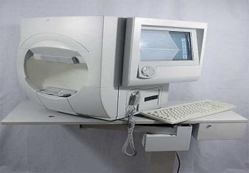
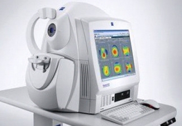
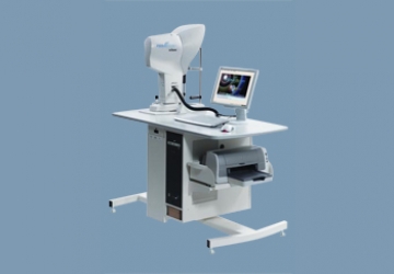
In the corneal topography test
In the corneal topography test, the refractive strength and the topographic structure of the cornea of the eye are measured, which makes up the bulk of the refractive power of the eye.
Read More..
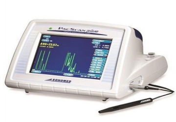
جهاز الموجات الصوتية واسعة المجال
يحتوي المركز على أحدث جهاز للتصوير بالموجات فوق الصوتية من انتاج شركة Sonomed الامريكية مزود بامكانيات فحص الجزء الأمامي من العين باستخدام مجسات خاصة ULTRASOUND BIOMICROSCOPY ، وتصوير الجزء الخلفي من العين لإعطاء تفاصيل عن الشبكية والجسم الزجاجي. كما يحتوي الجهاز على أحدث المعادلات العالمية لقياس قوة العدسة الخلفية والأمامية اللازمة للزرع داخل العين في حالات المياه البيضاء بدقة منقطعة النظير.
Read More..
جهاز التصوير المقطعي المحوسب الخاص بالعين OCT
جهاز التصوير المقطعي المحوسب الخاص بالعين OCT من شركة (TOPCON)اليابانية يعطي صور مقطعية دقيقة للشبكية لتشخيص أمراض الشبكية كاعتلال اللطخة الصفراء السكري و الشيخي و بعض الآفات الاخرى كثقوب مركز الإبصار كما يدرس هذا الجهاز بدقة متناهية العصب البصري وسماكة طبقة الالياف العصبية لتحديد مقدار تضرر العصب البصري نتيجة الاصابة بالزرق ( المياه السوداء) .
Read More..
جهاز تصوير قاع العين بأشعة الفلورسين
جهاز تصوير قاع العين بأشعة الفلورسين. وهو من انتاج شركة TOPCON اليابانية ويسمح بتصوير تفاصيل الجزء الخلفي من العين والشبكية ويمكن تصوير الدورة الدموية باستخدام تقنية FLUORESCEIN ANGIOGRAPHY كما توجد به خاصية تصوير العين INDOCYANINE GREEN ANGIOGRAPHY للتعرف على تفاصيل الأغشية المستحدثة أسفل مركز الإبصار.
Read More..
جهاز العلاج الضوئي الكيميائي (CORNEAL CROSS LINKING)
جهاز العلاج الضوئي الكيميائي (Corneal Cross Linking) وهي لتثبيت حالات القرنية المخروطية والأكتازيا.
Read More..
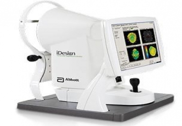
جهاز تصوير القرنية و دراسة الامواج الامامية
جهاز تصوير القرنية و دراسة الامواج الامامية (WaveScan Wave Front System) من شركة Abbot الامريكية وهو الجهاز الرائد لدراسة القرنية الانكسارية مما يسمح باجراء معالجة مفصلة لعين المريض مما يعطي افضل النتائج في تصحيح البصر بالليزر.
Read More..
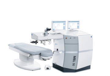
Excimer laser
Excimer laser
It is one of the latest scientific findings in treating the refractive defects of the eye, in which low-wave ultraviolet lasers are used to modify the surface of the cornea, and thus change its refractive strength. These rays remove layers of corneal tissue with pinpoint accuracy, with precisely defined length and depth.
Dr. Muhammad Hantira, an honorary assistant professor at the ophthalmology department, Umm Al-Qura University, Saudi Arabia, explained
That during the procedure of the laser there are those who monitor the process and watch it, through a computer located within the excimer laser tasked with determining the exact amount to be removed from the corneal tissue while maintaining the survival of the tissues soft and smooth.
How the laser works
Dr. Hantira explained:
The laser device works on shedding a beam of rays on the corneal tissue to be treated, which leads to those particles being dispersed from the cornea and thus changing its refractive power. In the event that these rays are shed inside the corneal tissue, it is called LASIK, which we will discuss in detail later. In the case of shining rays on the surface of the cornea, it is a superficial treatment, which is the subject of our discussion in this chapter.
The method of performing the operation
Dr. Hantira explained:
That, after the eye is given local anesthesia, the surface cells of the cornea (Epithelium) are removed manually, and then laser beams are applied to the outer surface of the cornea.
In order to speed up the healing of the outer layer of the corneal wound, and relieve the pain that some people may feel after the operation, a soft contact lens is placed, so that this lens is removed after the wound has healed, which may take 3 to 5 days.
It is worth noting that vision begins to improve gradually during the first weeks of the operation, but the final stability of vision may take 3 to 4 months.
It is one of the latest scientific findings in treating the refractive defects of the eye, in which low-wave ultraviolet lasers are used to modify the surface of the cornea, and thus change its refractive strength. These rays remove layers of corneal tissue with pinpoint accuracy, with precisely defined length and depth.
Read More..