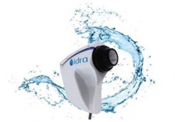
Idra Dry Eye diagnosis
Is the new instrument of individual analysis of tear film that allows to do a quick detailed structural research of the tear composition. Research on all the layers: Lipid, Aqueous, Mucin.
Thanks to I.C.P. IDRA it is possible to identify automatically the type of DED (Dry Eye Disease) and determine which layers can be treated with a specific treatment, in relation to the type of deficiency.
LIST OF AVAILABLE EXAMS
• AUTO INTERFEROMETRY
The IDRA automatically evaluates the quantity and quality of the lipid component on the tear film.
The device highlights the lipid layer and the software analyses automatically Lipid Layer Thickness (LLT).
• TEAR MENISCUS
The thickness of the tear meniscus that is observed on the eyelid margins provides useful information on the tear volume.
The tear meniscus can be examined considering its height, regularity and shape.
• NIBUT WITH MAP AND GRAPH
The stability of the mucin layer and the whole tear film is assessed through the study of the break up time (BUT)
Or non-invasive break up time (NIBUT), by using the Placido cone projected onto the cornea.
• MEIBOGRAPHY
Meibography is the visualization of the glands through illumination of the eyelid with infrared light.
It images the morphology of the glands in order to diagnose any meibomian gland drop out which would lead to tear dysfunction.
• 3D MEIBOGRAPHY
This new imaging system provides strong evidence to support the choice of a specific therapy (for example IPL treatment) and helps the patient to understand why a certain therapy is being recommended.
• BLINK RATE
It has been established that efficient blinking plays an important role in ocular surface health during contact lens wear and that it improves contact lens performance and comfort.
• BLEPHARITIS
This test helps to visually see blepharitis and presence of Demodex. It can be performed on the outer surface of the eye and eyelids.
• OCULAR REDNESS CLASSIFICATION
Once the image of the conjunctiva with its blood vessels is captured, it is possible to compare it with the classification sheets of bulbar and limbal redness degrees.
• PUPILLOMETRY
Measurement of the pupil reaction to light with and without glare. Measurement mode: SCOTOPIC, MESOPIC, PHOTOPIC
• WHITE TO WHITE MEASUREMENT
Evaluation of corneal diameter from limbus to limbus (white-to-white distance, WTW).