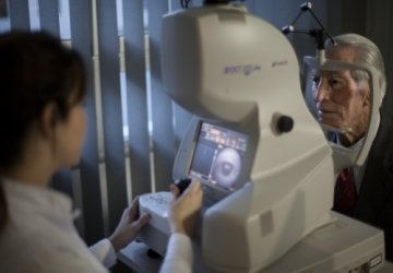
Optical coherence tomography (OCT) of the anterior segment
of the eye.
By asking Dr. Muhammad Hantira, Honorary Assistant Professor,
Ophthalmology Department, Umm Al-Qura University, Saudi Arabia,
about the role of the computerized tomography device in the field of
ophthalmology, he explained to us the following:
Optical coherence tomography (OCT) of the anterior segment of the
eye allows us to obtain high-resolution images from the front of the eye
using a property of light called optical interference. Currently it has
become a very useful tool for studying the eye's microscopic
anatomical structure.
Optical coherence tomography (OCT) of the anterior segment of the
eye uses:
• For follow-up of patients with refractive surgery, intra corneal
parenchyma, corneal transplantation, refractive cataract surgery.
• To follow up on patients who are undergoing surgery for glaucoma
leachate.
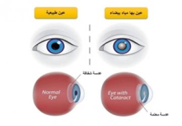
• In the field of cataract surgery, optical coherence tomography (OCT)
of the anterior segment of the eye allows accurate analysis of the
structure of the surgical incision, as well as the relationship between
the implanted lens and the posterior capsule.
Optical coherence tomography (OCT) of the anterior segment of the
eye is useful for:
• Analysis and evaluation of tumors and cysts in the front of the eye.
• Conjunctival tumors analysis, and various other diseases such as
corneal dystrophy, degeneration and infections.
• It also allows us to determine the thickness and epithelium of the
cornea.
• Determining the iris-corneal angle, measuring the depth of the
anterior chamber, and assessing the position of the implanted lens
inside the eye.
• In addition to studying the placing of contact lenses on the surface of
the cornea, and studying the condition of the surface of the eye, and it
is also useful for studying dry eyes.