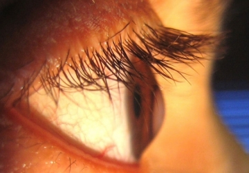
keratoconus
is a condition that affects the cornea of the eye and occurs when the cornea takes an abnormal shape and increases its curvature and protrudes outwards, turning from a spherical shape to a conical shape, due to the high pressure inside the eye.
Symptoms of keratoconus
Dr. Muhammad Hantira - Assistant Professor of the Ophthalmology Department - Umm Al-Qura University - Saudi Arabia explained
Keratoconus causes symptoms including:
- Vision gradually diminished in one or both eyes, often in late teens.
- Double vision when looking with one eye, even when wearing glasses.
- Seeing halos around bright lights.
- Glare and sensitivity to bright light which makes driving at night difficult.
- Blurry vision and gradually shift your vision.
- sudden changes in vision in one eye.
- Myopia and astigmatism.
Causes of keratoconus
Dr. Hantira explained:
The cause of keratoconus is unknown but genetic and environmental factors may contribute to the infection.
There are factors that increase the risk of developing keratoconus, including:
- A family history of keratoconus.
- Rub the eyes hard.
- Certain conditions such as retinitis pigmentosa, Ehler-Danlos syndrome, Down syndrome, hay fever, and asthma.
Diagnosis of keratoconus
Dr. Hanatira assured us That to diagnose keratoconus, the degree of curvature of the cornea must be measured, and that is done through the following tests:
- Ocular refraction
- Slit-lamp examination
- keratometry
- Computerized keratography
The latest laser treatment of keratoconus
Dr. Hantira explained to us:
The treatment of keratoconus depends on the stage in which the disease was discovered, as the development of keratoconus can be divided into 3 stages:
- The first (simple) stage: in which the vision is good with medical glasses
At this stage, keratoconus is fixed until the condition stabilizes so that no further deterioration occurs, as the surgeon uses compound drops of riboflavin placed on the surface of the cornea after removing the epithelial cells lining its front surface, and then UV rays are shed at specific doses that lead to an increase The bonds between the collagen fibers and the strengthening of the corneal tissue prevent deterioration for many years after this operation.
- The second (intermediate) stage: during which the patient's vision is not good with medical glasses.
At this stage, we treat keratoconus by implanting micro-rings inside the bark of the cornea using a femtosecond laser, which modifies the curvature and reduces the curvature of the cornea, which leads to a significant improvement in vision in most cases, then we fix keratoconus with ultraviolet rays and riboflavin so that no deterioration occurs one more time.
- The third stage (severe): in which the corneal opacities occur or a very severe curvature of the cornea.
At this stage, we treat advanced keratoconus, where we perform a corneal transplant (graft).