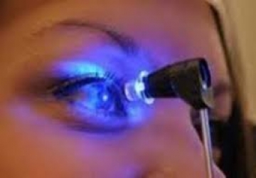
Intraocular presser
The term intraocular pressure refers to the pressure of the fluid inside the eye which is called aqueous humor.
This flow of fluids leads to the inability of the organs responsible for disposal to control them due to the accumulation of large amounts of it inside and outside the eye.
This leads to damage to the optic nerve, which is responsible for transmitting the image to the brain, and consequently, vision disturbance.
The intraocular pressure is responsible for giving the eye its circular shape, in addition to regulating the passage of nutrients and oxygen from the blood to the eye tissues.
intraocular pressure measurement
With the question of Dr. Muhammad Hantira - Honorary Assistant Professor - Department of Ophthalmology - Umm Al-Qura University - Saudi Arabia
He said that the pressure is considered normal if its ratio ranges between ten to twenty millimeters of mercury, and the eye suffers from eye pressure if its duration exceeds the normal limit.
It is measured using a precise device called a "tonometer" that measures the pressure of one or both eyes.
Symptoms of high intraocular pressure
Dr. Hantira explained:
It is difficult to detect or feel that eye pressure is high, but we present to you the most common symptoms:
• Feeling of sharp eye pain.
• severe redness of the eye.
• Feeling pain in the head.
• When looking directly at the light, the colors of the rainbow appear.
• Blurred vision.
• Vision becomes blurred as if the person is looking into a tunnel.
• Loss of vision.
• A blind spot in the external field of vision.
Candidates for high intraocular pressure
Dr. Hantereh advised:
It is advisable to regularly check your eye pressure for the cases below:
• People with diabetes and cardiovascular disease.
• Those who are obese.
• Those who are exposed to frequent eye infections.
• High blood pressure in the body.
• Those with cancerous and benign tumors inside the eye.
• Those over the age of sixty years.
How to diagnose high intraocular disease
When visiting Dr. Hanatera, he will conduct the following checks:
• Measuring with an instrument called a tonometer.
• Optic nerve examination.
• Visual field test, which is a test to assess peripheral vision.
• Examination of the angle course through which the fluids inside the eye are drained, where Dr. Hantereh evaluates the drainage system whether it is open or blocked.